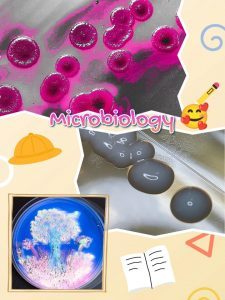
In the course of my work as a microbiologist, I’ve collected quite a bit of imagery. These are photos of cultivated microorganisms. Although difficult for a layperson to imagine, they are truly beautiful images that can be compared to abstract art. However, these are real living organisms that can sometimes take on strange yet very beautiful forms. I’ve digitally edited and artistically designed some of these photos. But microorganisms are also beautiful in their natural form.
Normally, most bacteria are invisible to humans because they are tiny (much, much smaller than 1 mm). But they are ubiquitous, meaning they are present everywhere: for example, in the environment, such as water or soil, but also on and in humans.
But how do you actually make bacteria and fungi visible?
Microorganisms can be cultivated in the laboratory or at home on nutrient media such as culture plates. To do this, you need to provide the tiny organisms with optimal conditions for reproduction. Culture plates consist of the basic substance agar agar and a host of important nutrients that bacteria and fungi need to grow. Additionally, you need to ensure a comfortable temperature. In medical microbiology, this is usually 35-37°C for bacteria.
Be inspired and discover the fantastic world of microorganisms
It’s fascinating to see how these tiny organisms develop on different culture media and the colors and shapes they take on. Some bacteria and fungi look almost like works of art, and it’s worth taking a closer look. The digitally enhanced photos often reveal a different side of the microorganisms, highlighting their beauty even more.
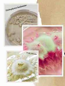
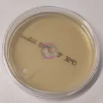
Trychophyton benhamiae
Trichophyton benhamiae is a dermatophyte found worldwide. These filamentous fungi cause specific fungal infections of the skin, known as dermatophytosis. T. benhamiae primarily causes dermatophytosis in children and adolescents, which is accompanied by a severe inflammatory skin reaction and intense itching. The infection manifests as bright red, centrifugally spreading, erythematous scales.
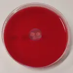
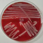
Staphylococcus aureus
Staphylococcus aureus belongs to the gram-positive cocci and is coagulase-positive. It can cause superficial and invasive purulent infections, as well as sepsis, pneumonia, and endocarditis. Several virulence factors, such as free coagulases, staphylokinase, and hemolysins, interact in its pathogenesis. Furthermore, some strains produce specific toxins, each responsible for vomiting and diarrhea, toxic shock syndrome (TSS), or staphylococcal-scalded skin syndrome (SSSS). Here you can see a unique growth pattern: S. aureus normally grows yellowish on CPSE agar, but in this case, it has a pleasing bluish hue.
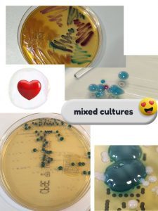
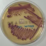
By the way, you can also paint with bacteria! Don't believe it? I'll show you how 🙂
It works best when using culture plates containing so-called chromogenic substances. These cause bacteria to grow in color on the plate. You then need a few plates on which bacteria have already grown. These are the equivalent colors you can choose from.
A loop serves as a brush, with which you apply the different colored bacteria to a culture plate. The difference from real painting is that you do it “blindly.” The result is only visible after about 24 hours, when the bacteria have multiplied and become visible.
Results
Here I tried to paint a tree, and I think I did quite well. I painted the tree trunk primarily with Escherichia coli, the treetop with Klebsiella spp., and the meadow with a touch of Pseudomonas aeruginosa. The pink and white touch in the flowers comes from both Staphylococcus saprophyticus and Staphylococcus epidermidis.
Baum nach 24h Bebrütung
Baum nach 48h Bebrütung
a few more examples
Just look at these beautiful pictures of bacteria and fungi.


